Dr. Shilpa Gandhi | Leading Consultant Minimally Invasive Thoracic Surgeon In Nagpur
Tracheal Stenosis

What is Tracheal Stenosis
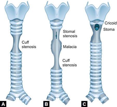
Your trachea, or windpipe, transports air from your nose and mouth to your lungs. Tracheal stenosis occurs when inflammation or scar tissue narrows the trachea, making breathing difficult.
There are two types of tracheal stenosis:
- Acquired tracheal stenosis: More common in children and adults, it develops due to illnesses or medical treatments affecting the trachea.
- Congenital tracheal stenosis (CTS): Present at birth, CTS is a rare and serious condition, affecting approximately 2 out of every 100,000 infants.
What are the symptoms of tracheal stenosis?
Symptoms of tracheal stenosis are similar in both children and adults. Common symptoms include:
- Difficulty breathing during everyday activities such as climbing stairs or walking.
- Wheezing.
- Persistent cough.
- Difficulty clearing mucus.
- Frequent respiratory infections like colds or pneumonia.
- Persistent asthma symptoms despite treatment.
- Chest congestion.
- Pauses in breathing (apnea) and sleep apnea.
Children may also experience specific symptoms:
- Infants may struggle with breastfeeding or bottle feeding and may appear excessively tired after feeding.
- Older children may have difficulty breathing or choking episodes while eating.
- The skin around the nose and gums of older children may appear blue.
- Both infants and older children may exhibit noisy breathing.
These symptoms vary in severity and may indicate tracheal stenosis, requiring medical evaluation and management.
How do healthcare providers diagnose tracheal stenosis?
How is tracheal stenosis treated?
Tracheal stenosis is typically managed with surgical interventions, chosen based on factors such as the degree of tracheal narrowing, its location, and the underlying cause. Common surgical treatments include:
Bronchoscopic tracheal dilation: Using a bronchoscope, a balloon or dilator is inserted into the trachea to expand the narrowed area, facilitating improved breathing. This procedure also provides detailed insights into the extent of tracheal narrowing.
Laser bronchoscopy: Employing a bronchoscope, a laser beam is directed at scar tissue within the trachea to alleviate obstruction.
Tracheal airway stent: A small tube made of plastic or metal is placed in the trachea to maintain its openness and ensure airflow.
Tracheal resection and reconstruction: In this procedure, the scarred or constricted portion of the trachea is surgically removed (resected), and the remaining healthy sections are sewn together to restore unobstructed airflow.


